Current Research

New Research article in Experimental Dermatology:
Digital Imaging Biomarkers Feed Machine Learning for Melanoma Screening
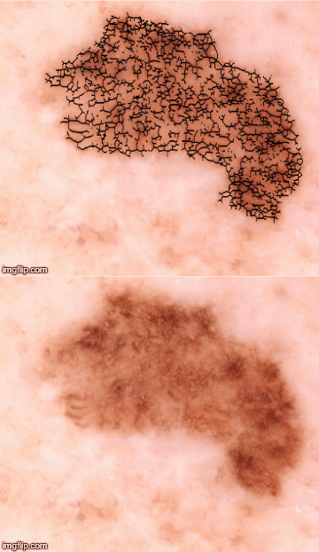
Automated detection of the pigmented network leads to quatitative metrics for melanoma screening. The link above will take you to the original article and the video below is a summary of the work.

Hi , I'm Dr. Dan Gareau, a biophotonic engineer. Having switched college studies from music (fuzzy logic) to physics, which is (mostly) universal, I migrated through electrical engineering (B.S. M.S.) to the LASER. Shining LASER light into dark corners of nature , I wonder: will cancer escape? Will we ever understand neuronal signaling? On a scale of 1 to 10, I like to turn the science up to eleven in the laboratory as a high-tech hacker, engineering devices that elucidate phenomena of critical importance.
One area of interest in my research is the use of confocal microscopy to image living cells and biological tissue. The video above is a chicken the beating heart of a chicken embryo. This is a perfect example of how LASERs and other optical components can be engineered into biological research tools. The videos below focus on the detection of cancer, a potentially life-saving event that can be achieves with advanced imaging.
Here is a talk I gave at the translational sciences conference:
The following projects are current works in progress in the lab:
 Hyperspectral Imaging for Melanoma Screening
Justin Martin, James Krueger, Daniel Gareau
The 5-year survival rate for patients diagnosed with Melanoma, a deadly form of skin cancer, in its latest stages is about 15%, compared to over 90% for early detection and treatment. We present an imaging system and algorithm that can be used to automatically generate a melanoma risk score to aid clinicians in the early identification of this form of skin cancer. Our system images the patient's skin at a series of different wavelengths and then analyzes several key dermoscopic features to generate this risk score. We have found that shorter wavelengths of light are sensitive to information in the superficial areas of the skin while longer wavelengths can be used to gather information at greater depths. This accompanying diagnostic computer algorithm has demonstrated much higher sensitivity and specificity than the currently commercialized system in preliminary trials and has the potential to improve the early detection of melanoma. Full Report (PDF)
 Automated Cellular Pathology in Noninvasive Confocal Microscopy
Monica Ting, James Krueger, Daniel Gareau
A computer algorithm was developed to automatically identify and count melanocytes and keratinocytes in 3D reflectance confocal microscopy (RCM) images of the skin. Computerized pathology increases our understanding and enables prevention of superficial spreading melanoma (SSM). Machine learning involved looking at the images to measure the size of cells through a 2-D Fourier transform and developing an appropriate mask with the erf() function to model the cells. Implementation involved processing the images to identify cells whose image segments provided the least difference when subtracted from the mask. With further simplification of the algorithm, the program may be directly implemented on the RCM images to indicate the presence of keratinocytes in seconds and to quantify the keratinocytes size in the en face plane as a function of depth. Using this system, the algorithm can identify any irregularities in maturation and differentiation of keratinocytes, thereby signaling the possible presence of cancer. Full Report (PDF)
Optical Fiber Spectroscopy Measures Perfusion of the Brain in a Murine Alsheimer’s Disease Model
Hyung Jin Ahn, Sidney Strickland, James Krueger, Daniel Gareau
Optical fiber spectroscopy is a versatile tool for measuring diffuse reflectance and extracting absorption information that can noninvasively quantify the presence of chromophores such as oxy-hemoglobin and deoxy-hemoglobin in tissues. Cerebrovascular abnormalities were widely recognized in Alzheimer’s disease (AD) patients. We analyzed blood volume fraction and level of oxygenated hemoglobin in Tg6799 mice, which are transgenic mice expressing five different familial Alzheimer disease-associated mutations in the human amyloid precursor protein and presenilin-1 genes. Diffuse reflectance spectra were iteratively fit as weighted sums of oxy- and deoxy-hemoglobin. Our observations showed slightly hypoxic conditions and significantly increased blood volume in the Alzheimer’s mice versus wild type. These results suggest that hyperperfusion of our AD mice may be a compensating mechanism for impaired cerebral vascular function and somehow relevant with early stage of AD patients. Ongoing work focuses on developing a cannula fixture that allows measurement in awake, behaving animals. Full Report (PDF)
 Raman Microbeam Spectrometer Noninvasively Measures Biomoelcules to Monitor the Tryptophan Metabolic Pathway
Gregory Michel, Jamie Harden, James Krueger, Alan Bigelow, Dan Gareau
Toward improving early detection of melanoma by accurate diagnosis and avoidance of unnecessary surgical excisions of common moles, we are developing noninvasive quantitative spectral fingerprinting of protein expression using Raman spectroscopy within confocally gated volumes of tissue. Our first target is the L-tryptophan catabolism pathway, which is unregulated in the tumor micro-environment and inhibits the immune response that usually is tumor suppressive. The tryptophan pathway is therefore worthy of diagnostic measurement and finding the ratio of L-tryptophan to its metabolites may aid a melanoma diagnosis. We report the intensity of the Raman signal from L-tryptophan and quinolinic acid, which are found during different stages of the tryptophan metabolic pathway. Full Report (PDF)
 Optical Properties of Cells With Melanin
Barukh Rhode, James Krueger, Daniel Gareau
The optical properties of pigmented lesions have been studied using diffuse reflectance spectroscopy in a noninvasive configuration on optically thick samples such as skin in vivo. However, it is difficult to un-mix the effects of absorption and scattering with diffuse reflectance spectroscopy techniques due to the complex anatomical distributions of absorbing and scattering biomolecules. We present a device and technique that enables absorption and scattering measurements of tissue volumes much smaller than the optical mean-free path. Because these measurements are taken on fresh-frozen sections, they are direct measurements of the optical properties of tissue, albeit in a different hydration state than in vivo tissue. Our results on lesions from 20 patients including melanomas and nevi show the absorption spectrum of melanin in melanocytes and basal keratinocytes. Our samples consisted of fresh frozen sections that were unstained. Fitting the spectrum as an exponential decay between 500 and 1100 nm [mua = A*exp(-B*(lambda-C)) + D], we report on the fit parameters of and their variation due to biological heterogeneity as A = 4.20e4 +/- 1.57e5 [1/cm], B = 4.57e-3 +/- 1.62e-3 [1/nm], C = 210 +/- 510 [nm] , D = 613 +/- 534 [1/cm]. The variability in these results is likely due to highly heterogeneous distributions of eumelanin and pheomelanin. Full Report (PDF)
 Monte Carlo Modeling of Pigmented Lesions
Daniel Gareau, Steven Jacques, James Krueger
Colors observed in clinical dermoscopy are critical to diagnosis but the mechanisms that lead to the spectral components of diffuse reflectance are more than meets the eye: combinations of the absorption and scattering spectra of the biomolecules as well as the “structural color” effect of skin anatomy. We modeled diffuse remittance from skin based on histopathology. The optical properties of the tissue types were based on the relevant chromophores and scatterers. The resulting spectral images mimic the appearance of pigmented lesions quite well when the morphology is mathematically derived but limited when based on histopathology, raising interesting questions about the interaction between various wavelengths with various pathological anatomical features. Full Report (PDF)
Anti-translational research: from the bedside back to the bench for reflectance confocal microscopy
Daniel Gareau
The reflectance confocal microscope has made translational progress in dermatology. 0.5 micrometer lateral resolution, 0.75mm field-of-view and excellent temporal resolution at ~15 frames/second serve the VivaScope well in the clinic, but it may be overlooked in basic research. This work reviews high spatiotemporal confocal microscopy and presents images acquired of various samples: zebra fish embryo where melanocytes with excellent contrast overly the spinal column, chicken embryo, where myocardium is seen moving at 15 frames/ second, calcium spikes in dendrites (fluorescence mode) just beyond the temporal resolution, and human skin where blood cells race through the artereovenous microvasculature. Full Report (PDF) also, see the poster version: Poster Link
 Multimodal confocal mosaics enable high sensitivity and specificity in screening of in situ squamous cell carcinoma.
Maria del Carmen Grados Luyando, Anna Bar Nicholas Snavely, Nathaniel Chen, Steven Jacques, Daniel S. Gareau
Screening cancer in excision margins with confocal microscopy may potentially save time and cost over the gold standard histopathology (H&E). However, diagnostic accuracy requires sufficient contrast and resolution to reveal pathological traits in a growing set of tumor types. Reflectance mode images structural details due to microscopic refractive index variation. Nuclear contrast with acridine orange fluorescence provides enhanced diagnostic value, but fails for in situ squamous cell carcinoma (SCC), where the cytoplasm is important to visualize. Combination of three modes [eosin (Eo) fluorescence, reflectance (R) and acridine orange (AO) fluorescence] enable imaging of cytoplasm, collagen and nuclei respectively. Toward rapid intra-operative pathological margin assessment to guide staged cancer excisions, multimodal confocal mosaics can image wide surgical margins (~1cm) with sub-cellular resolution and mimic the appearance of conventional H&E. Absorption contrast is achieved by alternating the excitation wavelength: 488nm (AO fluorescence) and 532nm (Eo fluorescence). Superposition and false-coloring of these modes mimics H&E, enabling detection of the carcinoma in situ in the epidermal layer The sum mosaic Eo+R is false-colored pink to mimic eosins’ appearance in H&E, while the AO mosaic is false-colored purple to mimic hematoxylins’ appearance in H&E. In this study, mosaics of 10 Mohs surgical excisions containing SCC in situ and 5 containing only normal tissue were subdivided for digital presentation equivalent to 4X histology. Of the total 16 SCC in situ multimodal mosaics and 16 normal cases presented, two reviewers made 1 and 2 (respectively) type-2 errors (false positives) but otherwise scored perfectly when using the confocal images to screen for the presence of SCC in situ as compared to the gold standard histopathology. Limitations to precisely mimic H&E included occasional elastin staining by AO. These results suggest that confocal mosaics may effectively guide staged SCC excisions in skin and other tissues. Full Report (PDF)